CASE DISCUSSION ON LIVER ABCESS
This is online E log book to discuss our patient's de-identified health data shared after taking his/her/guardian's signed informed consent. Here we discuss our individual patient's problems through series of inputs from available global online community of experts with an aim to solve those patients clinical problems with collective current best evidence based inputsThis e-log book also reflects my patient centered online learning portfolio and your valuable inputs on comment box is welcome.
Hyndavi Konakanchi , 8th semester
Roll no: 63
May 10, 2021
A CASE DISCUSSION ON LIVER ABCESS
I've been given this case to solve in an attempt to understand the topic of "patient clinical data analysis" to develop my competency in reading and comprehending clinical data including history, clinical findings, investigations and come up with a diagnosis and treatment plan.
Following is the view of my case:
(All this information provided by patient relative to Dr.Vamshi PG1)
Case: A 21 yr old male student,resident of nakrekal, came to the hospital with the CHEIF COMPLAINTS: of abdominal pain since 20 days and fever since 18 days
HISTORY OF PRESENT ILLNESS:
* Patient was apparently asymptomatic 20 days back and the he developed severe pain in epigastrium and right hypochondrium ( right upper quadrant) ,which was sudden in onset, gradually progressive ,and dragging type and radiating to right shoulder.
* There are no specific aggravating factors and releived upon medication and pain abdomen is not associated with nausea, vomiting and loose stools.
* And then later he developed Fever and loss of appetite since 18 days which was high grade and intermittent and associated with chills and rigor for 1 day and not associated with cold, cough, shortness of breath, headache, giddiness and vomiting.
* No history of chest pain, palpitations, burning micturition.
PAST HISTORY :
Patient was admitted in a local hospital in nakrekal, before with similar complaints 20 days back.There they counselled him regarding probable complications and need for referral for higher centre, So in advance he came to our hospital.
There is no history of Diabetes mellitus, Hypertension, Asthma and Epilepsy.
TREATMENT HISTORY:
Patient was given high dose IV antibiotics, antipyretics and analgesics
* Despite of which patient developed intermittent pain and fever and it also a reason for him to come to our hospital.
PERSONAL HISTORY:
Appetite: decreased since 18 days
Diet: Mixed
Bowel and bladder movements: Regular
No burning micturition
Addictions : Occasional toddy drinker
FAMILY HISTORY:
There is no significant family history.
GENERAL EXAMINATION:
The patient was conscious, coherent and cooperative, sitting comfortably on the bed.
He is well oriented to time , place and person.
He is thinly built and moderately nourished.
VITALS:
Temperature: Afebrile (at present)
Pulse: 82 beats per minute, regular , normal in volume and character. There is no radio radial delay and radio femoral delay.
Blood pressure: 110/70 mm of Hg
Respiratory rate: 14 cycles per minute
Jvp: normal
No Pallor
No Icterus
No Clubbing
No Lymphadenopathy
No Edema
( Vitals measured by Dr.Vamshi PG1)
SYSTEMIC EXAMINATION:
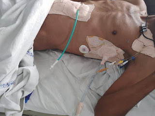
1) Abdominal examination:
Inspection:
Shape of abdomen: Flat / slightly scaphoid & symmetrical (normal)
(Picture mentioned below is captured by Dr. Vamshi PG1)

* Umbilicus : Normal ( inverted )
* No visible pulsations
* All quadrants of abdomen are moving equally on respiration.
Palpation:
* No local rise of temperature
* Tenderness is present over right hypochondrium and epigastrium
* No palpable masses foumd.
* Liver and spleen are not palpable
Percussion:
Liver span is normal (11 cm )
Auscultation:
Bowel sounds - heard
2)Respiratory system:
* Elliptical and bilaterally symmetrical chest
* Both sides moving equally with respiration
* Bi-lateral air entry — present
* Normal vesicular breath sounds
3)CVS:
* S1 and S2 heart sounds — heard
* No murmurs
( Examination done by Dr.Vamshi,PG1)
INVESTIGATIONS:
(Pictures of the data mentioned below are taken by Dr. Vamshi PG1)
Complete blood picture:
*leucocytosis

LFT ( Liver function tests ):
*Elevated ALP ( Alkaline phosphatase)

Renal function test(RFT):

Urine analysis:

Chest x-ray:
* Chest x-ray is done to rule out any respiratory pathologies
* In this case it is normal

Ultrasound(USG):
*Before treatment, A single oval shaped hyperechoic mass is seen in Right lobe of liver ( dated:3/5/21)
[ Identification of mass: is surrounded by markers ]

*After treatment, the mass became hypoechoic because of liquefaction of the mass ( ie in response to treatment)
1) In this usg image ; there is partial liquefaction (dated:6/5/21)

2) In this usg image ; there is 50-60% liquefaction (dated:8/5/21)

"The information below has been borrowed by me from various sources that I have referenced and acknowledged with their online links and I have paraphrased them further. However the images I have borrowed may have copyright issues as they may not be certified through a creative commons licence in which case I hope the original authors will get in touch with me and I shall remove them if they wish. I also request my readers to visit their original site as it was very useful to me."
Differential diagnosis:
LIVER- * Acute viral hepatitis
* Liver abcess
BILIARY- * Acute cholecystitis
*Acute cholangitis ( ascending cholangitis)
OTHERS- * Right lower lobe pneumonia
*Sub diaphragmatic abcess
Analysis of reports and diagnosis:
*Based on right hypochondriac and epigastric pain , fever
* USG finding of hyperechoic mass in right lobe of liver along with other supportive investigations like leucocytosis ( suggestive of infection/inflammation) and ALP ( Alkaline phosphatase ) rise in LFT is a suggestive diagnosis of LIVER ABCESS.
* Considering the following factors:
1) Age of the patient (21) - young & gender- male ,
2) Single abcess,
3) Right lobe involvement,
# The abcess is most likely to be AMOEBIC LIVER ABCESS.
* Since we cannot take risk , we should however administer antibiotics also ( like in pyogenic liver abcess)
TREATMENT ALONG WITH RATIONALE:
* INJECTION. ZOSTUM 1.5 gm IV BD (twice daily)
Zostum is a combination of drugs - SULBACTUM (pencillin) & CEFOPERAZONE(cephalosporin) [Antibiotic]: It is used here to treat if any bacterial cause ( since we can’t take the risk relying on only anti amoebic therapy)
* INJECTION. METROGYL 500mg IV TID ( thrice daily )
Metrogyl has the drug called METRONIDAZOLE [Antibiotic]: For amoebic cause
* INJECTION. OPTINEURIN 1amp in 100 ml NS( Nor
mal Saline) IV OD ( once daily)
Optineurin is a multivitamin drug { A combination of B1,B2, B3, B5,B6, B12 } given here as a supplement
* TAB. ULTRACET 1/2 QID( four times a day)
Ultracet is a combination of drugs - TRAMADOL(opiod analgesic) and ACETAMINOPHEN (analgesic and antipyretic) : Given for pain and fever
* TAB. DOLO 650 mg SOS (if needed) given for fever and pain
* Here ; due to medical therapy his symptoms subsided and clearly we can see it in usg reports ( liquefaction) meaning abcess responded to our medical therapy.
*And the patient was discharged on 10/5/21.
* We donot aspirate the pus since it is self resolving and aspiration is associated with several other complications.
* However there are some important indications for draining the abcess often asked in exams.
INDICATIONS OF LIVER ABCESS DRAINAGE:
1) If the abcess is large ( 5cm or more) because it has more chances to rupture.
2) If the abcess is present in left lobe as it may increase the chance of peritoneal leak and pericardial leak.
3) If the abcess is not responding to the drugs for 7 or more days
Method of abcess drainage:* Aspiration using needle or catheter under imaging
Reference:
https://emedicine.medscape.com/article/183920-overview-differential?src=mbl_msp_iphone
https://www.ncbi.nlm.nih.gov/books/NBK538230/
##If the patient has already had ruptured abcess ; immediate laparotomy and drainage of pus is done
https://images.app.goo.gl/xbp7Lr1dWRKPBAm86

TOPIC : LIVER ABCESS
* What is liver abcess ?
A liver abscess is defined as a pus-filled mass in the liver that can develop from injury to the liver or from an intra-abdominal infection disseminated from the portal vein.


* The majority of these abscesses are categorized as pyogenic or amoebic, although a minority are caused by parasites and fungi.
https://www.ncbi.nlm.nih.gov/books/NBK538230/
ETIOLOGY:
* Most amoebic infections are caused by Entamoeba histolytica. The pyogenic abscesses are usually polymicrobial, but some organisms are seen more commonly in them, such as E.coli, Klebsiella, Streptococcus, Staphylococcus, and anaerobes.
* While the incidence is low, it is essential to understand the severity of these abscesses because of the high mortality risk in untreated patients.
* If the cause is infectious, the majority of liver abscesses can be classified into bacterial (including amebic) and parasitic sources (including hydatiform cyst).
* The right lobe of the liver is more commonly affected than the left lobe.
https://www.ncbi.nlm.nih.gov/books/NBK538230/
RISK FACTORS:
* Poor personal hygiene.
* Malnutrition
* Use of steroids,diseases such as HIV (causes immunosuppression) and persons already having immunosuppression
* Heavily Crowded areas (slums)
* Men who have sex with men (secondary to sexually acquired amebic colitis)
https://emedicine.medscape.com/article/183920-overview-differential?src=mbl_msp_iphone
PATHOGENESIS:
* Since the liver receives its blood circulation from the systemic and portal circulations, it is more susceptible to getting infections and abscesses from the bloodstream.
* Proximity to gall bladder is another risk factor for the liver.
* The usual pathophysiology for pyogenic liver abscesses is bowel content leakage and peritonitis. Bacteria travel to the liver via the portal vein and resides there. Infection can also originate in the biliary system. Hematogenous spread is also a potential etiology.
* Septic emboli cause several microabscesses which combine to form one large abscess.

* Hematogenous spread from endocarditis or pyelonephritis can happen.
https://www.ncbi.nlm.nih.gov/books/NBK538230/
CLINICAL FEATURES:
History:
* Clinical features are non specific for liver abcess and are similar to other febrile diseases.
* There is no difference in clinical presentation in both amoebic and pyogenic abcess.
Time of onset:
*The most common complaints being fever and abdominal pain in any liver abcess.
*In amoebic liver abcess this type of clinical presentation is more common in young male patients.
*And coming to the duration of symptoms it may vary anywhere between 2 weeks.
Abdominal pain:
* This is the first most common complaint received from patient and present in almost all patients.
*Type of pain: Constant and dull aching in right hypochondriac and epigastric regions , which may radiate to right shoulder or scapular regions due to common nerve innervation.
## Aggrevating and relieving factors: The pain increases with coughing, walking, and deep breathing, as well as when patients rest on their right side.
Constitutional symptoms:
* These include FEVER,NAUSEA&VOMITING,DIARRHOEA
* Fever is almost present in all cases.
* In some cases there are pulmonary symptoms ( cough , chest pain ) indicating the ruptured abcess in pleural cavity.
https://emedicine.medscape.com/article/183920-overview-differential?src=mbl_msp_iphone
Physical examination: ( signs)
*The most common sign: Fever (99% cases)
*Next to that tenderness in Right upper quadrant (right hypochondrium) and in some cases tenderness in epigastrium is noted.
* In some cases: Hepatomegaly and jaundice are also present.
Type of jaundice: OBSTRUCTIVE JAUNDICE, being if the abcess is large / multiple,it : they compresses the biliary tract.
https://emedicine.medscape.com/article/183920-overview-differential?src=mbl_msp_iphone
INVESTIGATIONS:
Hematology:
*Common hematological finding being LEUCOCYTOSIS , indicating inflammation or infection.
* Anemia may be present , or not.
Chemistry:
* Elevated liver enzymes such as AST,ALP are noted .
* Similar reports are found in both amoebic liver abcess and pyogenic liver abcess.
* Hyperbilurubinemia is present in small no.of cases where the jaundice is present.
https://emedicine.medscape.com/article/183920-overview-differential?src=mbl_msp_iphone
Stool examination, culture , Antigen detection:
## But most patients with amoebic liver abscess have no bowel symptoms, examination of stool for ova and parasite and antigen testing is insensitive and not recommended.
* And Considering the risk factors associated with the aspiration of pus for culture:
1) Some times abscess is not accessible for aspiration if it is in posterior aspect or so
2) Sometimes it has thinwall which may rupture if u aspirate
3) Sometimes it is unliquified
##Then how can u confirm whether it is pyogenic/amoebic , so we treat both emperically in clinical practice.
Imagining studies:
Ultrasonography:
*It is preferred because it is rapid , inexpensive and sensitive compared to other costly procedures.
* And also it has an advantage of avoiding radiation exposure.
https://emedicine.medscape.com/article/183920-overview-differential?src=mbl_msp_iphone
* For example; notice in the below image 3 well defined, rounded lesions are seen in usg.

CT SCAN, MRI:
* These are sensitive but not specific and also the information provided by them is almost same as other inexpensive imaging methods.
* So these are not commonly used.
https://emedicine.medscape.com/article/183920-overview-differential?src=mbl_msp_iphone
TREATMENT:
* As mentioned earlier in practice we treat both pyogenic and amoebic liver abcess empirically.
* So we cover both bacterial causes with broad spectrum antibiotics and also amoebic causes mostly with metronidazole.
* Next we administer patient with analgesic and antipyretic such as tab.dolo 650mg &tab.Ultracet , to releive pain and fever.
##Abcess may get ruptured if untreated and cause peritonitis and shock.

Comments
Post a Comment